1  DICOM Basic

1.1 Overview of DICOM

A DICOM file (Digital Imaging and Communications in Medicine) is a standardized file format used to store medical imaging data, along with detailed metadata that describes the image and other related information. The DICOM standard ensures that images and related data are consistently structured, allowing for interoperability across different imaging devices and medical systems (e.g., PACS). Here’s a breakdown of how data and metadata are organized in a DICOM file:

1.1.1 1. File Structure Overview

Each DICOM file is structured into two main components:

  • Header (Metadata): Contains information about the image, the patient, the acquisition settings, and other related details.

  • Pixel Data (Image): The actual image data (or voxel data in the case of 3D scans like MRI or CT) is stored here.

1.1.2 2. DICOM Header (Metadata)

The DICOM header contains metadata that describes the image and provides context. This metadata is organized into Data Elements, which are composed of:

  • Tag: A unique identifier for each element, written as a hexadecimal number (e.g., (0010,0010) for the patient’s name).
  • Value Representation (VR): Specifies the data type for the value (e.g., text, integer, float).
  • Length: Indicates the size of the data element in bytes.
  • Value: The actual information being stored (e.g., the patient’s name, scanner settings, slice location, etc.).

1.1.2.1 Common Metadata Categories:

  • Patient Information:
    • Patient’s name, ID, birth date, sex (Tags like (0010,0010) for patient’s name).
  • Study Information:
    • Study date, description, referring physician, etc. (e.g., (0008,0020) for Study Date).
  • Series Information:
    • Series number, series description, modality type (e.g., MRI, CT).
  • Image Acquisition Parameters:
    • TR, TE, flip angle, slice thickness, b-values (for DWI), and other imaging parameters.
  • Image Information:
    • Slice location, pixel spacing, image orientation, image dimensions (e.g., (0028,0010) for the number of rows in the image, (0028,0011) for the number of columns).

1.1.2.2 Example of DICOM Header:

(0010,0010) - Patient's Name - John Doe
(0010,0020) - Patient's ID - 12345
(0008,0020) - Study Date - 2023-09-01
(0008,103E) - Series Description - Brain MRI
(0020,0013) - Image Number - 5

1.1.3 3. Pixel Data (Image Data)

The actual medical image data (whether 2D or 3D) is stored in the Pixel Data element, identified by tag (7FE0,0010). This part contains the raw image data, which can be structured in different formats (grayscale or color, compressed or uncompressed).

1.1.3.1 Characteristics of Pixel Data:

  • Pixel dimensions: Defined by metadata tags, such as:
    • Rows (0028,0010): The number of rows in the image.
    • Columns (0028,0011): The number of columns in the image.
  • Pixel Spacing: The physical distance between pixels (defined by tag (0028,0030)).
  • Bits Allocated: Describes the bit depth (the number of bits used per pixel, typically 8, 12, or 16 bits for grayscale medical images).
  • Rescale Slope and Rescale Intercept: Used to convert raw pixel data into meaningful units (e.g., Hounsfield Units in CT scans).

1.1.4 4. Data Organization in a DICOM File

The data in a DICOM file is organized in Data Sets, which follow a hierarchical structure. Each Data Set contains Data Elements. Data Elements are composed of: - Tag: The unique identifier for that piece of data (like a label). - Value Representation (VR): The type of data (e.g., Integer, String, Float, etc.). - Value: The actual data, such as patient information, image dimensions, etc.

1.1.5 Example of DICOM Data Element:

(Tag)      (VR)      (Length)   (Value)
(0010,0010) PN        8          John Doe
(0010,0020) LO        6          12345
(0008,0020) DA        8          20230901

In this example: - (0010,0010): Tag for “Patient Name” (PN). - (0010,0020): Tag for “Patient ID” (LO – Long String). - (0008,0020): Tag for “Study Date” (DA – Date).

1.1.6 5. Handling 3D or Multislice Data (Volumes)

For volumetric imaging like CT or MRI, multiple DICOM files are typically generated, where each file corresponds to a single slice of the entire volume. Each file contains: - Its own header describing slice location, thickness, and orientation. - The pixel data for that specific slice.

Software like PACS or 3D Slicer will aggregate these individual files and combine the slices to create a full 3D volume for viewing or analysis.

1.1.7 6. Encapsulation and Compression

DICOM allows for encapsulating images in different formats and compressing pixel data (e.g., JPEG or JPEG 2000). The choice of compression is noted in the metadata, ensuring that systems can correctly decode the data.

1.1.8 7. DICOM Metadata Example in a Simple MRI Scan

Here’s a simplified example of what you might see in a brain MRI DICOM file:

  • Patient’s Name: John Doe (0010,0010)
  • Modality: MR (0008,0060)
  • Slice Thickness: 5 mm (0018,0050)
  • Pixel Spacing: 0.9 mm x 0.9 mm (0028,0030)
  • Image Orientation: Specifies how the image is oriented in space (0020,0037)
  • Rows and Columns: 512 x 512 (0028,0010 and 0028,0011)
  • Pixel Data: Contains the actual image data (7FE0,0010)

1.1.9 8. DICOM File Size

The size of a DICOM file varies depending on:

  • Image resolution: Higher resolution means more pixel data.
  • Number of slices: For volumetric sequences (like MRI or CT), each slice generates a separate DICOM file.
  • Bit depth: More bits per pixel (e.g., 16-bit vs. 8-bit) increases the file size.
  • Compression: Use of lossy or lossless compression affects the size of the pixel data.

1.1.10 Summary

  • DICOM Header (Metadata): Contains descriptive data such as patient information, study parameters, and image acquisition details.
  • Pixel Data (Image Data): Stores the actual image data for a slice, organized as a matrix of pixel intensity values.
  • Multiple Files per Volume: For 3D scans like MRI or CT, each slice has its own DICOM file, and these files are combined by the viewing software to create a complete 3D volume.

This structure makes DICOM files both versatile and complex, allowing them to carry detailed metadata along with high-quality medical images.
1.2 DICOM as Python Obj

To represent DICOM files from a single MRI sequence as a Python data structure, you can organize the metadata and pixel data using a list of dictionaries, where each dictionary represents a single DICOM file (or slice) in the MRI sequence. Each dictionary would contain keys for metadata fields (such as patient information, acquisition parameters, and image-specific details) and pixel data stored in a NumPy array (which is often how medical images are processed in Python).

Here is an example of a Python data structure representing multiple DICOM files in a single MRI sequence:

Code
import numpy as np

# Example DICOM sequence with metadata and pixel data for each slice
mri_sequence = [
    {
        'FileName': 'slice_001.dcm',
        'PatientName': 'John Doe',
        'PatientID': '123456',
        'StudyDate': '2023-09-01',
        'SeriesDescription': 'Brain MRI - T1WI',
        'SliceLocation': -40.0,
        'SliceThickness': 5.0,
        'PixelSpacing': [0.9, 0.9],
        'ImageOrientationPatient': [1, 0, 0, 0, 1, 0],
        'Rows': 512,
        'Columns': 512,
        'PixelData': np.random.randint(0, 4096, (512, 512), dtype=np.uint16)
    },
    {
        'FileName': 'slice_002.dcm',
        'PatientName': 'John Doe',
        'PatientID': '123456',
        'StudyDate': '2023-09-01',
        'SeriesDescription': 'Brain MRI - T1WI',
        'SliceLocation': -35.0,
        'SliceThickness': 5.0,
        'PixelSpacing': [0.9, 0.9],
        'ImageOrientationPatient': [1, 0, 0, 0, 1, 0],
        'Rows': 512,
        'Columns': 512,
        'PixelData': np.random.randint(0, 4096, (512, 512), dtype=np.uint16)
    },
    # ... more slices
]

# Example of accessing data from one DICOM slice
first_slice = mri_sequence[0]
print("Patient Name:", first_slice['PatientName'])
print("Slice Location:", first_slice['SliceLocation'])
print("Pixel Data Shape:", first_slice['PixelData'].shape)
Patient Name: John Doe
Slice Location: -40.0
Pixel Data Shape: (512, 512)

1.2.1 Explanation of Data Structure:

  1. List of Dictionaries: Each dictionary in the mri_sequence list represents a single DICOM file or slice in the MRI sequence.
  2. Metadata:
    • FileName: The file name of the DICOM slice.
    • PatientName: The patient’s name.
    • PatientID: Unique ID for the patient.
    • StudyDate: Date of the MRI study.
    • SeriesDescription: Description of the MRI series (e.g., “T1WI”).
    • SliceLocation: The z-coordinate of the slice relative to some reference point.
    • SliceThickness: The thickness of the slice.
    • PixelSpacing: The distance between adjacent pixels, typically in millimeters.
    • ImageOrientationPatient: Specifies the orientation of the image slice relative to the patient.
    • Rows and Columns: The resolution of the image slice.
  3. PixelData:
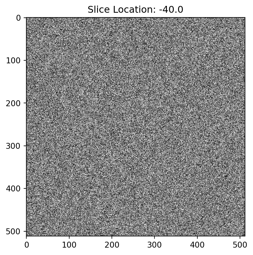
    • This is a NumPy array that holds the actual pixel intensity values for that slice. Each slice in the MRI sequence has its own pixel data. Here, I’ve used np.random.randint to simulate pixel values in the range of 0–4096, which is typical for 12-bit medical images.

1.2.2 Accessing and Processing:

You can easily access the pixel data for each slice and perform operations such as:

  • Viewing the shape of the image slice (first_slice['PixelData'].shape).
  • Performing image processing using libraries like NumPy or SciPy.
  • Using visualization tools like matplotlib to display the slices.

1.2.3 Example of Displaying the First Slice:

Code
import matplotlib.pyplot as plt

# Display the first slice
plt.imshow(mri_sequence[0]['PixelData'], cmap='gray')
plt.title(f"Slice Location: {mri_sequence[0]['SliceLocation']}")
plt.show()

This data structure is a Pythonic way to represent DICOM files in an MRI sequence, making it easy to store both metadata and image data in a structured format. You can then loop through the list to access individual slices, process pixel data, or export them into different formats.